


The different types of ocular imaging examinations
To provide an accurate diagnosis and the right treatment pathway that’s adapted to each type of eye disease, eye doctors can choose to prescribe ocular imaging examinations. These examinations are offered in addition to standard visual acuity tests.
Optical coherence tomography
Optical coherence tomography, or OCT, is based on the same principle as echography, with the difference that it uses light rather than ultrasound. It enables the retina to be observed and measured from a cross-sectional view.
This examination is indispensable for the diagnosis of diseases like AMD or diabetic macular edema, as well as for thorough monitoring and follow-up.
A non-invasive procedure that involves no direct contact with the eye, optical coherence tomography sometimes requires prior dilation of the pupil using eye drops.
Retinography
Retinography consists in taking a photograph of the posterior part of the eye, notably the retina. This type of snapshot enables the ophthalmologist to detect certain retinal diseases (AMD, diabetic macular edema…) with more precision.
This exam can be carried out with or without prior dilation by an ophthalmologist or orthoptist.
Angiography
Angiography is a procedure that is used to produce images of blood vessels – in many different parts of the body, including the eyes. This technique involves tracking the movement of a contrast dye which is first injected into the blood vessels, in order to better visualize the vascularization of the retina.
This procedure can facilitate the confirmation of diagnosis of AMD and of diabetic retinopathy, and it can also be used to guide the delivery of certain treatments, such as laser photocoagulation.
In case of dilated pupils, ask someone to accompany you, or opt to take public transport, as this can disturb your vision for several hours.
 |
|
LOW VISION GLASSES
For patients with AMD.
|
 |
|
SETTINGS
Performed by a health professional. |
 |
|
CUSTOMER SERVICE
By email by |

Sign up to be notified when the glasses are available:
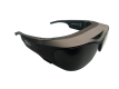

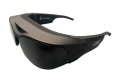
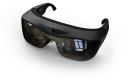


Light Vision Glasses for AMD patients
Light Vision develops glasses with eyetracking technology and control softwarefor AMD patients to improve their daily lives and give them back some autonomy.
FOLLOW-US!